
Dahms Lab – Dept Chemistry and Biochemistry – U Regina
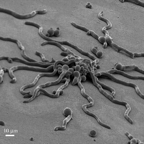
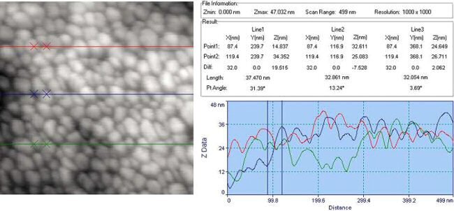
Maturation of the hyphal wall of Aspergillus nidulans using atomic force microscopy (AFM) and cryo-scanning electron microscopy (cryoSEM eg.). These complementary methods image cell surfaces in frozen hydrated (cryoSEM) or vapour-fixed critical point dried samples (SEM and AFM). AFM has superb xyz resolution. CryoSEM preserves lifelike spatial relationships and has excellent xy resolution. We are now using AFM to image the surface of growing hyphae.
This AFM image was collected by Ms. Hui Ma:
This cryoSEM was taken with the assistance of George Braybrook, Scanning Electron Microscope Lab, Dept Earth & Atmospheric Sciences, U Alberta: